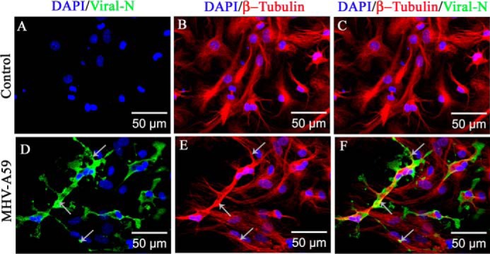
Figure 3.

Association of MHV-A59 virus particles with microtubules in primary astrocytes. Primary astrocytes were infected with MHV-A59 at an MOI of 2, and mock-infected cells were maintained in parallel. Cells were subjected to double-label immunofluorescence with rabbit anti-β-tubulin antiserum (red) and mouse anti-N antiserum (green). Cells were counterstained with DAPI (blue), and merged image projection (mip) signals were imaged with an apotome microscope. No viral N–specific signal was observed in mock-infected cells (A), but viral N staining was observed to be dispersed from the perinuclear space to the surface of MHV-A59–infected cells (D, arrow). MT morphology is shown in mock-infected (B) and virus-infected (E, arrow) cells. MHV-A59–infected cells showed the virus-specific signal colocalizing with the MTs. Specifically, near the cell periphery, viral N signal was present on MT threads (F, arrow). As expected, no viral N signal was seen in mock-infected cells (C).
