
Figure 6.
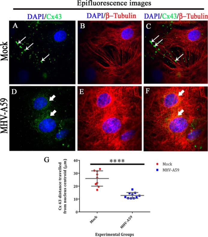
Whole-cell expression of Cx43 and β-tubulin upon MHV-A59 infection. Primary astrocytes immunolabeled for Cx43 and β-tubulin, which was subjected to TIRF microscopy, were simultaneously taken for epifluorescence microscopy to obtain the whole-cell Cx43 expression. Thus, parallel epifluorescence images were captured for the same field. Cx43 was observed to be present in profuse amounts as its characteristic punctate stain of Cx43 (A (thin arrow) and C (merged)). In contrast, MHV-A59–infected astrocytes showed mainly perinuclear localization of Cx43 (D (thick arrow) and F (merged)), which was not observed by TIRF imaging. MT morphology is shown for mock-infected (B) or MHV-A59–infected cells (E). The distance of Cx43 molecules from the nucleus (distance was calculated from nuclear centroid) was measured with the help of ImageJ (G). For mock-infected cells, Cx43 was present ∼25.9 μm away, which was reduced to ∼12.7 μm in MHV-A59–infected cells. Data were obtained from nine different images from n = 3 biological replicates, and average ± S.D. (error bars) is represented (****, p < 0.0001; t test).