
Figure 2.
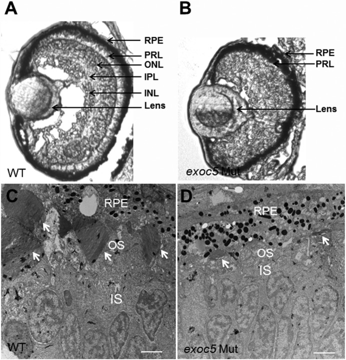
Histologic analysis of retinas of wild-type and exoc5 mutant zebrafish larvae. A and B, transverse sections of 3.5-dpf WT (A) and exoc5 mutant (B, exoc5 Mut) eyes. In exoc5 homozygous mutants, retinal lamination was lost, and the photoreceptor outer segments (arrows; photoreceptor layer (PRL)) were disorganized and not readily detectable. C and D, ultrastructural analysis of WT and exoc5 mutant photoreceptors using transmission electron microscopy. C, WT photoreceptors showed tightly stacked disc membranes (arrows) and inner segments; D, in exoc5 mutants only remnants of outer segments (arrows) could be observed. Scale bars, 2 μm (C and D). OS, outer segments; IS, inner segments; ONL, outer nuclear layer; RPE, retinal pigmented epithelium.