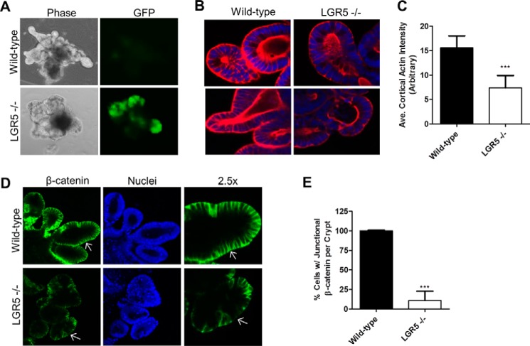
Figure 1.
Loss of LGR5 in intestinal crypt organoids resulted in disorganization of F-actin and β-catenin. A, bright-field micrographs of wild-type and LGR5 KO mouse intestinal crypt organoids cultured in Matrigel. B, confocal microscopy images of F-actin (red) in wild-type and LGR5 KO organoids. Nuclei were counterstained using TO-PRO-3 (blue). C, quantification of cortical F-actin. D, confocal images of β-catenin in WT and LGR5 KO organoids. Nuclei were counterstained using TO-PRO-3 (blue). Arrows are included for image reference. E, quantification of β-catenin. Error bars are S.D. (n = 15–20 crypts). ***, p < 0.001 versus WT.

