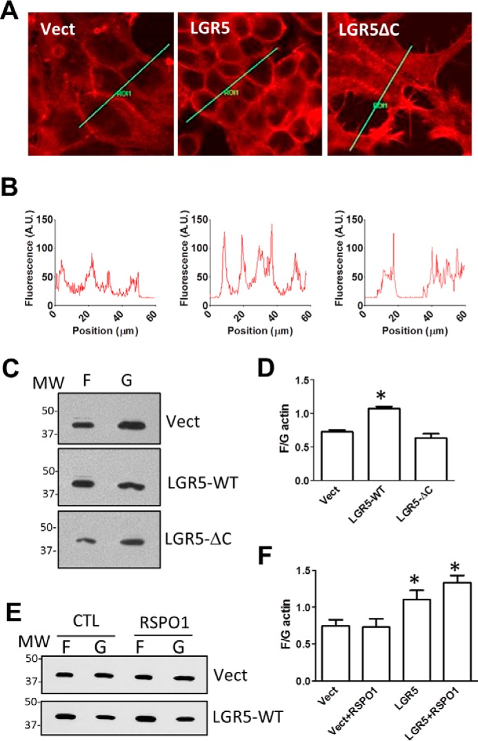
Figure 3.

Overexpression of LGR5 in HEK293T cells increased cortical F-actin levels. A, confocal images of phalloidin staining of HEK293T cells expressing vector control, Myc-tagged LGR5-WT, or LGR5-ΔC. B, quantification of fluorescence of the lines drawn in A. C, representative WB results of G- and F-actin as separated by the Triton X-100 method. D, quantification of F- and G-actin WB results expressed as the ratio of F- versus G-actin. E, representative WB results of G- and F-actin of HEK293-vector or -LGR5-WT cells after treatment with RSPO1 (100 ng/ml) or vehicle overnight. F, quantification of WB results of G/F-actin. All error bars are S.E. (n = 3). *, p < 0.05 compared with vector (Vect) cells. A.U., arbitrary units; CTL, control.
