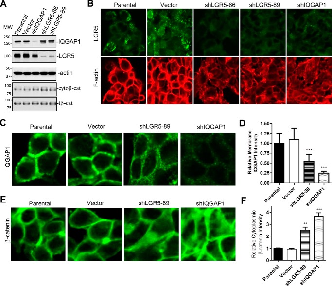
Figure 7.
KD of LGR5 in LoVo colon cancer cells altered cytoskeletal structure and decreased the level of membrane-associated IQGAP1 and β-catenin. A, WB results in control and LoVo cells with KD of IQGAP1 or LGR5. B, confocal images of LGR5 (green) in control and cells with KD of IQGAP1 or LGR5. F-actin was stained with rhodamine-labeled phalloidin (red). C, confocal images of IQGAP1 in control and LoVo cells with KD of LGR5 or IQGAP1. D, quantification of membrane-associated IQGAP1. Error bars are S.E. (n = 20–30 cells). ***, p ≤ 0.001 versus parental and vector cells. E, confocal images of β-catenin. F, quantification of cytoplasmic β-catenin based on relative signal intensity. Error bars are S.E. (n = 20–30 cells). ** and ***, p ≤ 0.01 and 0.001, respectively, compared with parental and vector cells. Images in C and E are 2.5× magnification compared with B. cytoβ-cat, cytoplasmic β-catenin; tβ-cat, total β-catenin.

