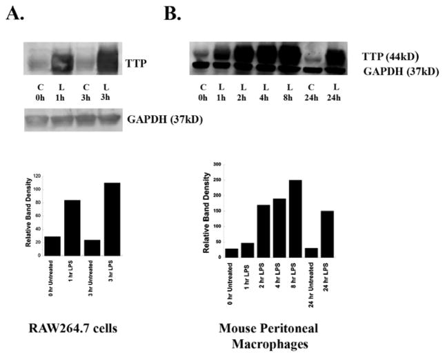
Figure 4. LPS induces Expression of TTP in RAW264.7 Cells and Murine Peritoneal Macrophages (MPMs).
RAW264.7 cells and MPMs from C57Bl/6J mice were treated with LPS (100ng/ml), or with RPMI-1% FBS alone as control (C) for various time periods. Following treatment, media were removed, cells were solubilized in SDS-polyacrylamide gel electrophoresis sample buffer and lysates (50μg protein/sample) were Western-blotted with TTP antibody. GAPDH was used as a loading control. Each experiment was performed twice with similar results. A typical Western blot for each cell type is shown. A: RAW264.7 cells. B: Mouse peritoneal macrophages. Band intensities were quantified by using ImageQuant analysis program (GE Health care).

