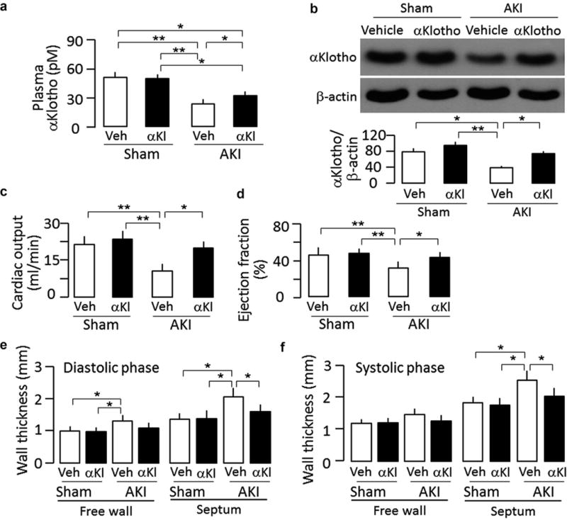
Figure 1. αKlotho administration after acute kidney injury (AKI) maintained higher plasma and renal αKlotho levels and improved cardiac function in ischemia-reperfusion injury (IRI)–induced AKI mice.

Sham-treated or IRI-induced AKI wild-type mice were Injected i.p. with αKlotho (αKl) protein (0.01 mg/kg) or vehicle (Veh) (phosphate-buffered saline) for 4 consecutive days starting 24 hours after surgery. Throughout the experimental period, all mice were fed normal rodent chow. At 20 weeks after surgery, animals were killed. (a) Plasma αKlotho. (b) αKlotho protein in the kidney. Upper panel shows representative immunoblots for αKlotho and (β-actin in the kidney. Bottom panel is a summary of normalized protein quantification from all examined immunoblots. (c) Cardiac output, (d) Left ventricular ejection fraction, (e) Left ventricular wall thickness at diastole, (f) Left ventricular wall thickness at systole. Data are expressed as means ± SD of 4 mice from each group, and statistical significance was assessed by 1-way analysis of variance followed by Student-Newman-Keuls test, and accepted when: *P < 0.05, **P < 0.01 between 2 groups.
