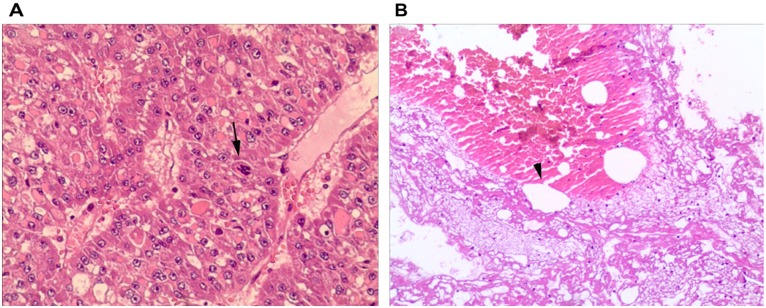
Figure 3.
Pathological examination of liver specimens from clamping and non-clamping groups demonstrated: (A) Disordered hepatic cords, dark, multipolar and heteromorphic nuclei and mitose atypique identifying moderate differentiation hepatocellular carcinoma (arrow), (hematoxylin and eosin; magnification, ×100); (B) discontinuity of small venous wall, venous rupture and thrombi at the site of rupture (hematoxylin and eosin; magnification, ×400).

