Figure 1.
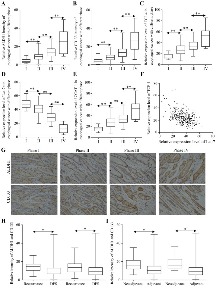
The correlation between clinicopathological patterns and signatures of stem cell enrichment in esophageal cancer. The expression levels of ALDH1 (A) and CD133 (B) in esophageal cancer of different stages were detected by immunohistochemistry, and the differences between ratios of stem cells are significant. The relative expression levels of TCF4 (C), Let-7b (D) and CCAT1 (E) in esophageal cancer of different stages were detected by qRT-PCR, and the difference between the expression of each stage is significant. (F) The correlation between levels of TCF-4 and Let-7b was analyzed with Pearson correlation coefficient, results showing R=−0.4621, P<0.0001, 95% confidence interval: −0.5445 to −0.3708. (G) The representative immunohistochemistry images for ALDH1 and CD133 intensity in esophageal cancer are shown. The expression levels of ALDH1 and CD133 in patients with 2-year recurrence or disease-free survival are presented (H), and the different expression levels of ALDH1 and CD133 in patients who underwent adjuvant or neoadjuvant chemotherapy were identified (I). *P<0.05, **P<0.01.

