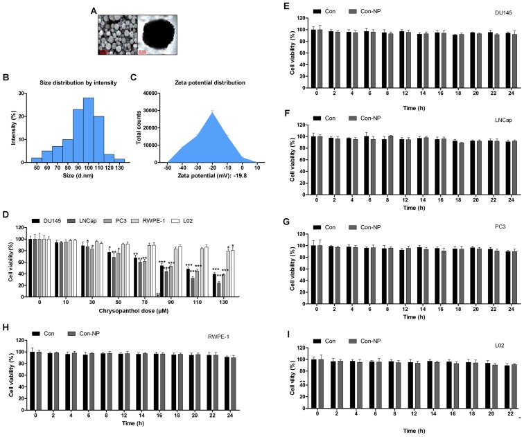
Figure 1.
Gold-chrysophanol nanoparticle characterization and its cytotoxicity to prostate cancer cells. (A) The scanning electron microscopy (SEM) images of chrysophanol nanoparticles. (B) Dynamic light scattering (DLS) image of cerium oxide nanoparticles. (C) Zeta potential of PLGA encapsulated chrysophanol nanoparticle. (D) Chrysophanol nanoparticle (0–130 μM) was added to human prostate cancer cell line cultures and human prostate normal epithelia cells, as well as normal liver cell culture for 24 h. The cell viability was calculated by MTT assay, and the graph exhibits a gradual downregulation in prostate cancer cell viability. (E) DU145, (F) LNCap, (G) PC3, (H) RWPE-1, and (I) L02 cells were not given any treatment (Con) or only the nanoparticles (Con-NP) for different times as indicated (0, 2, 4, 6, 8, 12, 14, 16, 18, 20, 22 and 24 h). Then, the cell viability was evaluated using MTT analysis. Data are shown as mean ± SEM. *P<0.05, **P<0.01 and ***P<0.001 versus the control group in the absence of any treatment.

