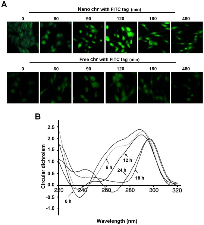
Figure 4.
The interaction of chrysophanol nanoparticle with DNA of prostate cancer cells. (A) LNCap cells were treated with 90 μM chrysophanol nanoparticle (upper) or free chrysophanol (lower) for 0–480 min. The cells were visualized and analyzed at the described time intervals under a fluorescence microscope. Greenish cell fluorescence refer to the presence of chrysophanol nanoparticle inside the cancer cell. (B) DNA from LNCap cells was extracted at the indicated time intervals. The interaction between chrysophanol nanoparticle and DNA was investigated by circular dichroism (CD) spectroscopy. Data are shown as mean ± SEM.

