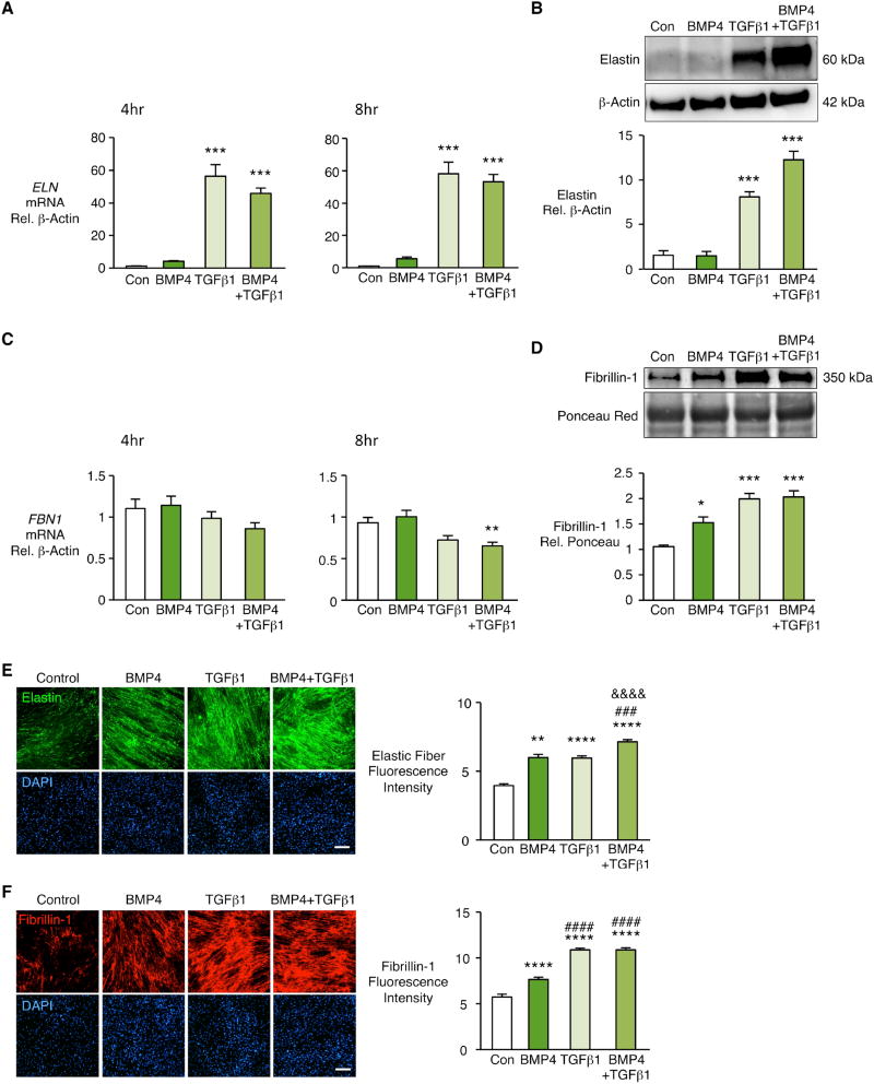
Figure 1. TGFβ1 increases elastin mRNA and protein and TGFβ1 and BMP4 increase extracellular fibrillin-1 in human pulmonary artery fibroblasts.
Human pulmonary artery fibroblasts (PAF) isolated from unused donor lungs and used between passages 3–6 were stimulated with BMP4 (10ng/ml), TGFβ1 (2ng/ml), BMP4+TGFβ1, or vehicle (Con). (A, C) Fold change in mRNA of ELN and FBN1 was measured four and eight hours after stimulation. (B, D) Representative immunoblots above and densitometry below for elastin in cell lysates and fibrillin-1 in conditioned media 48h after stimulation. Beta-actin and Ponceau staining were used as loading controls for cell lysates and conditioned media respectively. Fibrillin-1 is normalized to Con. Bars represent Mean±SEM of n=3–5 independent experiments, *p<0.05, **p<0.01, ***p<0.001 vs. Con, by one-way ANOVA and post-hoc Bonferroni test.
(E, F) PAF of donor controls were seeded in glass chamber slides and grown for four days to confluence. Cells were starved overnight, then stimulated every other day for seven days with vehicle (Con), BMP4, TGFβ1 or BMP4+TGFβ1. Elastic fibers were visualized by indirect immunofluorescence of elastin (E) and fibrillin-1 (F). Right, fluorescence intensities quantified by Image J software and normalized to cell number assessed by nuclei DAPI staining. Scale bar=100μm. Bars represent Mean±SEM of n=4 independent experiments, **p<0.01, ****p<0.0001 vs. Con; ###p<0.001, ####p<0.0001 vs. BMP4; &&&&p<0.0001 vs. TGFβ1, by two-way ANOVA and post-hoc Bonferroni test.

