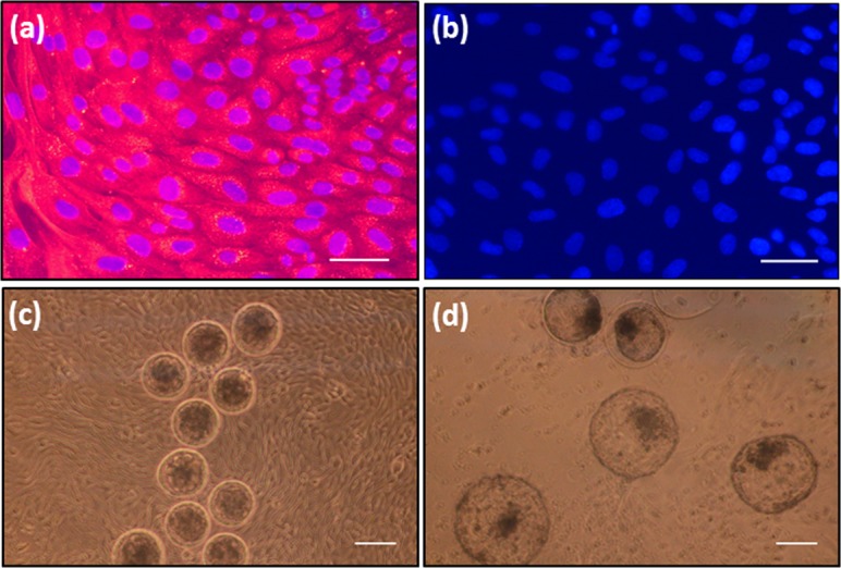
Fig. 2.

(a) Immunofluorescence image of cultured BUECs with anti-cytokeratin antibody. Goat anti-rabbit IgG Alexa Flour 546 (red) was the secondary antibody, DAPI (blue) was used to visualize nuclei (b) Immunofluorescence image of cultured BUECs without anti-cytokeratin antibody as negative control. PBS-T (0.1% Tween-20 in PBS–/–) was used instead of primary antibody (c) Morulae on the BUEC monolayer at the start of 4 day co-culture, and (d) Blastocysts on the BUEC monolayer at the end of 4 day co-culture. Scale bar = 100 µm for a, b and 200 µm for c, d.
