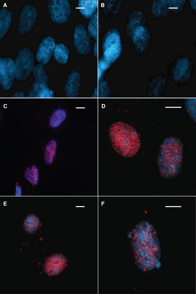
Figure 4.
Cellular localization of FANCA protein. Red, FANCA; blue, DAPI. Nuclear localization of null FANCA control showing no FANCA signal with (A) no mitomycin C (MMC) and (B) 1 mM MMC. Nuclear localization of wild-type FANCA with (C) no MMC and (D) 1 mM MMC. Nuclear locatization of FANCA in the S1088F FANCA cell line with (E) no MMC and (F) 1 mM MMC. Scale bar, ∼10 µm.

