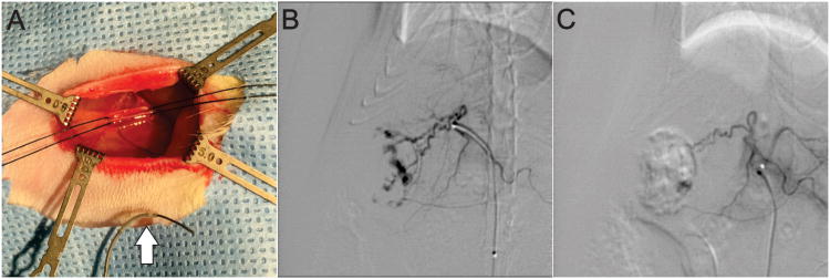
Figure 5.
(A) Intra-operative photo demonstrating the surgical field for the transfemoral arterial approach. 5-0 silk sutures can be seen positioned around the femoral artery as vessel loops. A 1.5F catheter with guidewire can be seen in the foreground for scale (white arrow). (B) Pre-embolization selective arteriogram showing superselective catheterization of the artery feeding the superior right lobe of the liver. (C) Post-embolization selective arteriogram demonstrating nearly complete embolization of large tumor as indicated by reduced contrast filling.

