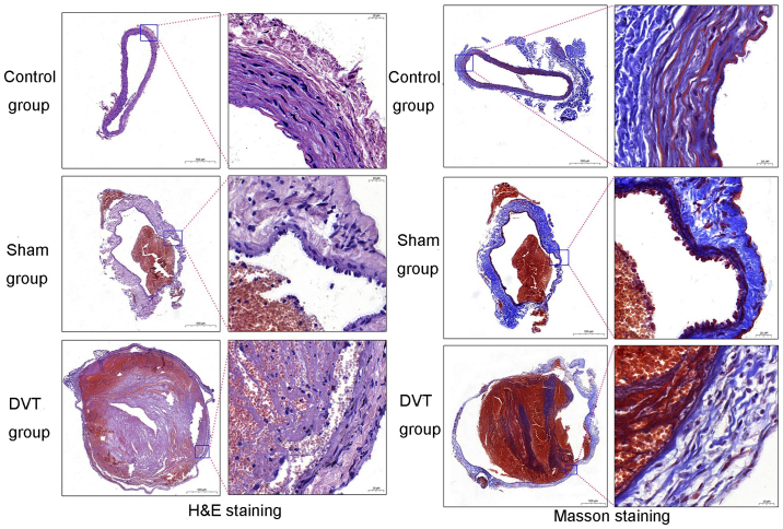
Figure 2.
Photomicrographs of the sections stained with H&E and Masson's trichrome. H&E and Masson staining exhibiting normal inferior vena cava in the control group. Accumulation of blood cells is apparent in the veins of the sham group, while no fibrin is observed. No thrombosis is present in the control and sham groups. Vascular endothelial cells in the DVT group are not continuous, and the images exhibit substantial numbers of inflammatory cells infiltrating and surrounding the blood vessels. The thrombus in the lumen is a mixed thrombus. Collagen fiber hyperplasia occurred and partially entered the thrombus. H&E, hematoxylin and eosin; DVT, deep vein thrombosis. Scale bars of the left pictures of H&E or Masson staining represents 500 μm; scale bars of the right pictures represents 20 μm.

