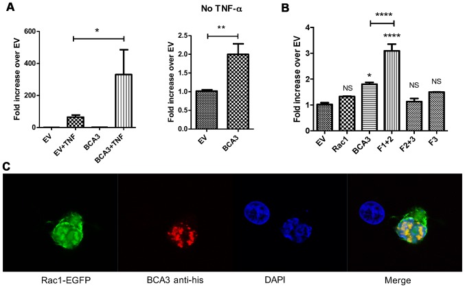
Figure 2.
Breast cancer associated gene 3 (BCA3) augments nuclear factor-κB (NF-κB) signaling and co-localizes with Rac1 in 293 cells. (A) An NF-κB luciferase reporter construct and a BCA3 expression vector were co-transfected into 293 cells. Luciferase assays demonstrated that BCA3 significantly increased NF-κB activity after tumor necrosis factor-α (TNF-α) treatment (left panel; 10 ng/ml for 24 h). The right panel shows the effect of BCA3 overexpression on NF-κB signaling in the absence of TNF-α and is simply the data from columns 1 and 3 in the left panel presented on a different scale so the effect can be seen. Data were analyzed by an unpaired t-test. (B) Rac1, full length BCA3 or the indicated truncated BCA3 constructs were co-transfected with the NF-κB luciferase reporter into 293 cells. BCA3 fragment 1+2 augmented NF-κB signaling to a significantly greater extent than did either full-length BCA3 or fragment 2+3. Data were analyzed by one-way ANOVA with Bonferroni post-test corrections. (C) Vectors containing Rac1-EGFP and His-tagged, full-length BCA3 were co-transfected into 293 cells, stained and imaged by confocal microscopy. BCA3 and Rac1 co-localized primarily in the nucleus of 293 cells (*p<0.05; **p<0.01; ***p<0.001; NS, p>0.05). NS, not significant.

