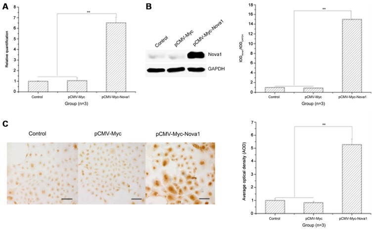
Figure 1.
The mRNA and protein expression levels of neuro-oncological ventral antigen 1 (Nova1) in PC12 cells transfected with pCMV-Myc-Nova1 as detected by RT-qPCR, western blotting and cell immunocytochemistry. (A) The mRNA expression level of Nova1 as detected by RT-qPCR. (B) The protein expression level of Nova1 as detected by western blotting. (C) The protein expression and distribution of Nova1 as detected by cell immunocytochemistry. The images indicate that at 48 h after transfection, the mRNA and protein expression levels of Nova1 were significantly increased to a plateau, and widely distributed in the cytoplasm and nuclei (**p<0.01). Scale bar, 100 µm.

