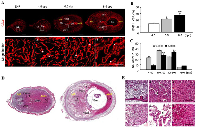
Figure 1.
Changes (vascular remodeling) occurring in the endometrium during pregnancy. (A) Images showing CD31+ BVs in the uterus at ENP, 4.5, 6.5 and 8.5 days post coitum (dpc). Scale bars, 500 µm. Each numbered magnification image (square-dotted line) shows endometrial CD31+ BVs in the VSR. Scale bars, 100 µm. (B) Comparisons of CD31+ BV densities (BVD, %) in the VSR at 4.5, 6.5 and 8.5 dpc. Each group, n=5–6. *p<0.05 vs. 4.5 dpc; **p<0.01 vs. 4.5 dpc by unpaired t-test. (C) Comparisons of numbers of different sized BVs in the MR at 6.5 and 8.5 dpc. Each group, n=5–6. *p<0.05 vs. 6.5 dpc; **p<0.01 vs. 6.5 dpc by unpaired t-test. (D) Cross-sectioned uterus from 6.5 to 8.5 dpc stained with hematoxylin and eosin. Scale bars, 500 µm. (E) Magnified images showing enlarged and elongated vascular lumen (arrowheads) in the uterus at 6.5 to 8.5 dpc. Scale bars, 100 µm. MR, mesometrial region; AMR, anti-mesometrial region; UL, uterus lumen; VSR, venous sinus region; Em, embryo; BVs, blood vessels.

