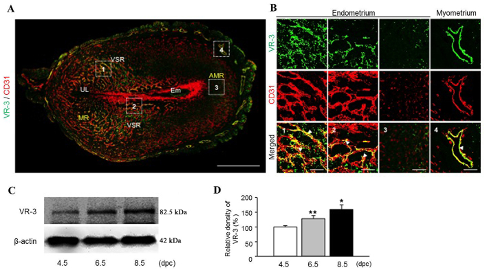
Figure 2.
Vascular endothelial growth factor receptor-3 (VEGFR-3) expression increases during pregnancy in the uterus. (A) Image showing CD31+ BVs and VEGFR-3+ cells in the uterus at 6.5 days post coitum (dpc). Scale bars, 500 µm. (B) Magnified images showing VEGFR-3 expressed CD31+ BVs (arrowheads) in the endometrium and myometrium of uterus at 6.5 dpc. Scale bars, 50 µm (C) Comparison of VEGFR-3 protein levels in the uterus at 4.5 to 8.5 dpc. Levels of VEGFR-3 were normalized to those of β-actin. (D) The VEGFR-3 protein levels in the uterus at 4.5 to 8.5 dpc as shown in (C). Each group, n=5–6.*p<0.05 vs. 4.5 dpc; **p<0.01 vs. 4.5 dpc by unpaired t-test. VR-3, VEGFR-3; MR, mesometrial region; AMR, anti-mesometrial region; UL, uterus lumen; VSR, venous sinus region; Em, embryo.

