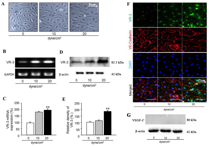
Figure 4.
Fluid shear stress regulates vascular endothelial growth factor receptor-3 (VEGFR-3) expression in human uterine microvascular endothelial cells (HUtMECs). (A) Morphology of HUtMECs following 24 h of exposure to static (0 dyne/cm2) and fluid shear stress (FSS) (10–20 dyne/cm2) condition. (×100 magnification). (B) Comparison of VEGFR-3 mRNA expression in HUtMECs after 24 h exposure to static (0 dyne/cm2) and FSS (10–20 dyne/cm2) conditions. Levels of VEGFR-3 were normalized to GAPDH. (C) The vascular endothelial growth factor receptor-3 (VEGFR-3) mRNA levels in human uterine microvascular endothelial cells (HUtMECs) as described in (B). **p<0.01 vs. controls (0 dyne/cm2) by unpaired t-test. (D) Comparison of VEGFR-3 protein expression in HUtMECs after 24 h exposure to static (0 dyne/cm2) and fluid shear stress (FSS) (10–20 dyne/cm2) conditions. Levels of VEGFR-3 were normalized to β-actin. (E) The VEGFR-3 protein levels in HUtMECs as described in (D). **p<0.01 vs. controls (0 dyne/cm2) by unpaired t-test. (F) Representative immunocytochemistry of HUtMECs after 24 h exposure to static (0 dyne/cm2) and FSS (10–20 dyne/cm2) conditions to determine VEGFR-3 and VE-Cadherin protein levels. Scale bars, 100 µm. (G) Comparison of VEGF-C protein expression in HUtMECs after 24 h exposure to static (0 dyne/cm2) and FSS (10–20 dyne/cm2) conditions. Levels of VEGF-C were normalized to those of β-actin.

