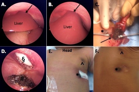
Figure 2: A and B. Laparoscopic images showing the left lateral segment of the liver in continuity with the external portion of the mass. The black arrow demonstrated the liver exiting through the chest wall. The left hemidiaphram was significantly elevated. C. Electrocautery was used to amputate the mass where it bordered the normal liver (black arrow). D. Post amputation of the exophytic mass demonstrated a circular chest wall defect (§), and hemostatic liver edge (#). E. Fascial closure and skin closure without further defect noted. Normal (^) and accessory left nipple (*) F. Three-month follow up demonstrating well-healed incision and mild chest wall deformity.
