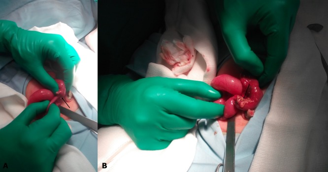
Figure 1: A: Intraoperative view showing the ileo-ileal intussusception (arrow). B: Intraoperative view, after reduction, showing the perforated Meckel’s diverticulum (arrow)

Figure 1: A: Intraoperative view showing the ileo-ileal intussusception (arrow). B: Intraoperative view, after reduction, showing the perforated Meckel’s diverticulum (arrow)