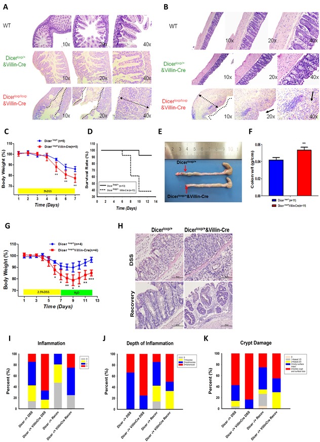
Figure 1. Dicer heterozygous mice are prone to DSS induced colitis.

A.-B. H&E staining the small intestine (A) or large intestine (B) of WT mouse (upper panel), Dicerloxp/+&VillinCre mouse (middle panel), and Dicerloxp/loxp&VillinCre mouse (bottom panel) respectively. C.-F. Dicerloxp/+ mice (n = 5, blue) and Dicerloxp/+&VillinCre mice (n = 5, red) were used in acute colitis model. Body weight (C) results and survival days (D) were recorded; the length of the intestine (E) and the colon weight length radio (F) are presented. Statistical significance was determined using a two-tailed, unpaired Student’s t-test; P < 0.01. G.-K. Dicerloxp/+ mice (n = 4, blue) and Dicerloxp/+&VillinCre mice (n = 4, red) were used in sub-acute colitis mouse models. Mice body weight (G) was recorded. Morphology of the intestine was observed by H&E staining (H) (magnification, × 400). Score of the inflammation degree (I), the depth of inflammation (J) and crypt damage (K) was conducted.
