
Figure 2. Dicer deletion impairs the stable status of cells in vivo and in vitro.
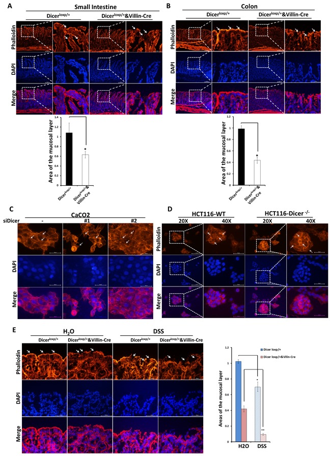
A.-B. Rhodamine phalloidin staining experiments were performed using frozen sections of small intestine and colon tissues from Dicerloxp/+ mice and Dicerloxp/+&VillinCre mice. Representative images for Small intestine (A, Upper panel) and Colon (B, Upper panel) are shown (magnification, left×100, and right × 400). Quantitation of the mucosal area (magnification, × 400) are shown in (A, Bottom panel) and (B, Bottom panel) for small intestine and colon respectively. C.-D. Caco2 cells transfected with siDICER siRNAs for 48h (C) and HCT116-WT and HCT116-DICER−/−cells (D) were stained with Rhodamine phalloidin. Representative images are shown (magnification, × 400). E. Dicerloxp/+ mice and Dicerloxp/+&VillinCre mice were sacrificed and colon tissues were taken for rhodamine phalloidin staining (left). Representative images are shown (magnification, × 400). Quantitation of the mucosal layer is shown (right). Nuclei were all stained with DAPI (Blue).