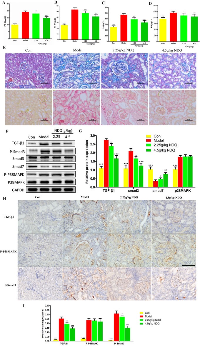
Figure 7. Effects of NDQ on fibrosis-related indices and signaling pathways.

A.-D. Four fibrosis-related indexes (PCIII, COLIV, LN, and FN) were measured by ELISA. E. Histopathological findings in rat remnant kidneys analyzed on week 19. Renal tissue sections were stained with Masson's trichrome stain for interstitial fibrosis and Sirius red stain for collagen fibers (200×, scale bar represents 500 μm). F. Rat kidneys were harvested at the end of the experiment, and analyzed to identify the expression of TGF-β1, Smad2/3, p-Smad2/3, Smad7, p38MAPK, and p-p38MAPK by western blotting. GAPDH served as the loading control. G. Densitometric analysis of TGF-β1, Smad2/3, smad7, and p38MAPK expression. H. TGF-β1, p38MAPK, and Smad3 expression in the remnant kidney of CKD rats were analyzed by immunohistochemistry (200×, scale bar represents 500 μm). I. Expression of TGF-β1, p-p38MAPK, and p-Smad3 was analyzed quantitatively. Data are presented as means ± SD. *p < 0.05, **p < 0.01, ***p < 0.001 (versus untreated model group).
