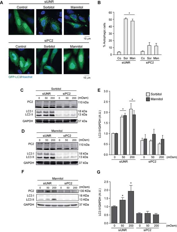
Figure 3. PC2 is required for hyperosmotic stress-induced autophagy.

PC2 was downregulated in HeLa cells by a specific siRNA against PC2. An unrelated siRNA (UNR) was used as a control. Subsequently, cells were infected with AdGFP-LC3 for 24 h and treated with sorbitol or mannitol (200 mOsm) for 2 h. Cells were fixed, and autophagy was evaluated by fluorescence microscopy. Representative pictures are shown in A. The percentage of autophagic cells is shown in B. (mean ± SEM, n = 3, *p < 0.05 vs. Co siUNR). Nuclei were dyed with 10 ng/mL of DAPI C.-G. PC2 was downregulated in HeLa C.-E. and HCT116 F.-G. cells with a specific siRNA against PC2. LC3 I-to-LC3 II conversion was evaluated by Western blot analysis in HeLa C.-E. and HCT116 F.-G. cells exposed to sorbitol or mannitol (0, 50 or 200 mOsm) for 2 h. Representative gels are shown in C., D. and F.. Quantification of gel bands is shown in shown in E. and G. (mean ± SEM, n = 3, *p < 0.05 vs. 0 mOsm siUNR). GAPDH levels were used as a loading control.
