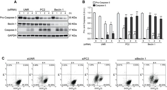
Figure 4. Pro-survival role of autophagy in cells subjected to hyperosmotic stress.

HeLa cells were transfected with an unrelated control siRNA (siUNR) or specific siRNAs against PC2 and Beclin 1. 48 h later, cells were exposed to sorbitol (200 mOsm) at the indicated times. Pro-caspase-3, Beclin 1 and caspase-3 levels were evaluated by Western blot analysis. GAPDH levels were used as a loading control. Representative gels are shown in A. Quantification of gel bands is shown in B. (mean ± SEM, n = 3, *p < 0.05 vs. 0 h siUNR, **p < 0.01 vs. 0 h siUNR). Alternatively, cells were submitted to cytofluorimetric analysis of mitochondrial membrane potential (DiOC6(3) staining) and viability (PI staining). C. Representative dot plots of HeLa cells treated for 6 h with 200 mM sorbitol are shown (n = 3).
