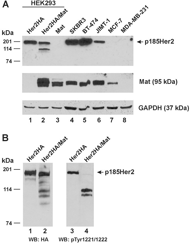
Figure 5. Relative expression of Her2 and matriptase in experimental cell lines and cleavage of phosphorylated Her2 by matriptase.

(A) An equal amount of total protein lysate (20 μg) from HEK293 cells transfected with Her2HA (Lane 1), Her2HA and matriptase (Mat, Lane 2), or matriptase (Mat, Lane 3), and the human breast cancer cells SKBR3 (Lane 4), BT-474 (Lane 5), JIMT-1 (Lane 6), MCF-7 (Lane 7), and MDA-MB-231 (Lane 8) was used for western blot analysis of Her2 (Top Panel) and matriptase (Middle Panel). A GAPDH western blot (Bottom Panel) was performed as a loading control. (B) HEK293 cells transfected with Her2HA (Lanes 1 and 3), or Her2HA and matriptase (Mat, Lanes 2 and 4) were analyzed for Her2(HA) (Left Panel) and phospho-Her2 (pTyr1221/1222) (Right Panel). WB: western blot.
