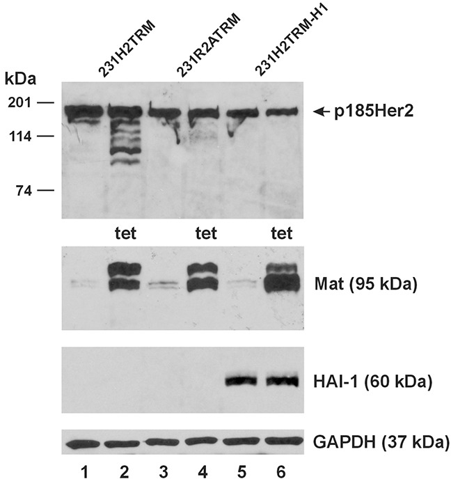
Figure 6. Matriptase cleaves stably expressed Her2 but not Her2 with mutations at the matriptase cleavage sites.

An equal amount of total protein lysate (40 μg) from the 231H2TRM (Lanes 1 and 2), 231R2ATRM (Lanes 3 and 4), and 231H2TRM-H1 (Lanes 5 and 6) cells was used for western blot analysis of Her2HA (Top Panel), matriptase (Mat, Second Panel from the Top), and HAI-1 (Third Panel from the Top). Cells represented by samples in Lane 2, 4, 6 were treated with 1 μg/ml tetracycline (tet) for 24 hours. A GAPDH western blot (Bottom Panel) was performed as a loading control.
