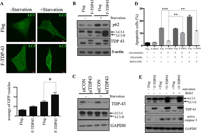
Figure 3. TDP-43 promotes autophagy formation under nutrient deprivation.

(A) Autophagosome formation in starved cells. U87MG cells stably over-expressing TDP-43 or Flag control were transfected with GFP-LC3 and were grown in either complete medium or nutrient-deprived medium. Autophagosome formation was analyzed by flourescent microscopy. Quantitative data are expressed as the numbers of GFP punctates in a cell (mean±SD, lower). (B and C) Western blotting of LC3 expression. Cells starved for indicated times were subjected to Western blotting using P62, LC3 and TDP-43 specific antibodies. β-actin was used as a loading control. (D) Apoptotic cells under nutrient deprivation. U87MG cells stably expressing TDP-43 or Flag control were treated with BafA1 (0.5 nM) or 3-MA (1 mM), respectively, and were cultured with or without starvation for 24 hours. After starvation, cells were stained with annexin V/PI and measured by flow cytometry. (E) Western blotting of active caspase 3 in U87MG. Cells treated with DMSO, Bafa1 (0.5 nM) were starved for the 24 hours, and were collected and subjected to Western blotting using LC3, TPD-43, Caspase-3 specific actibodies. GAPDH was used as internal control.
