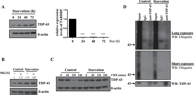
Figure 4. TDP-43 degradation is inhibited in nutrient deprivation condition.

(A) TDP-43 expression under starvation. 05MG cells were starved for indicated time and total cell lysates were subjected to Western blotting using TDP-43 specific antibody. β-actin was used as a loading control. The mRNA level of TDP-43 under starvation. U87MG cells starved for indicated time were collected and total RNA was analyzed by quantitative PCR. 18S was used as internal control. (B) Protein expression of TDP-43 under starvation condition. 05MG cells were nutrient-deprived for 8 hours with or without 25uM MG132. Expression of TDP-43 in total cell lysates was analyzed by Western blotting using an anti-TDP-43 antibody. β-actin was used as a loading control. (C) U87MG cells were treated with DMSO or CHX (50 um) for indicated time in starvation (HBSS) or complete medium. TDP-43 expression was analyzed by immunoblotting using TDP-43 specific antibody. β-actin was used as a loading control. (D) Ubiquitination of TDP-43 in starved cell. Total cell lysates from nutrient-deprived cells or control cells were immunoprecipitaed with TDP-43 antibody and subjected to Western blotting. The signals were detected by ubiqunitin or TDP-43 specific antibody.
