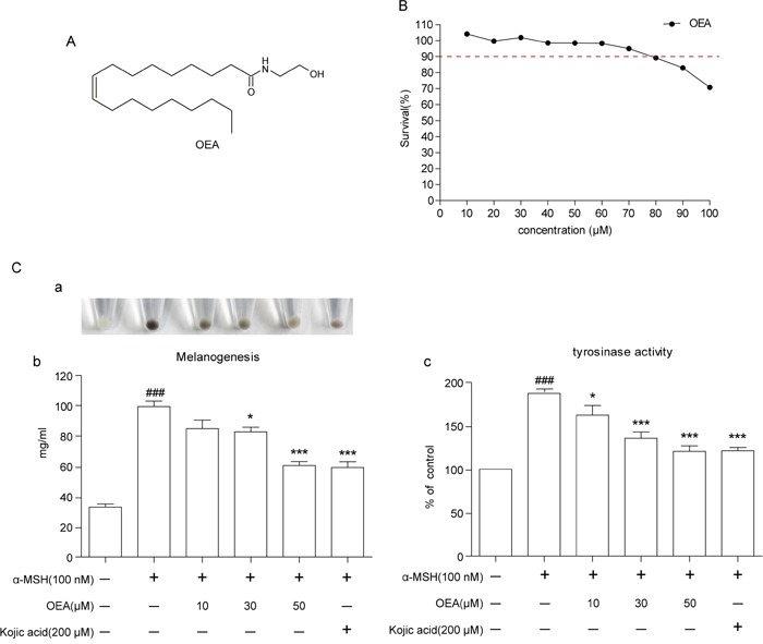
Figure 1. Effect of OEA on cellular melanin synthesis and tyrosinase activity in α-MSH-stimulated B16 cells.

(A) The chemical structure of OEA. (B) Cells were treated with various concentrations of OEA for 72 h. Cell viability was determined by MTT assay. (C) Relative cellular melanin content and tyrosinase activity were measured at 72 h after treatment. Cells were exposed to 100 nM α-MSH in the presence of 10 to 50 μM OEA or 200 μM kojic acid. The percentage values of the treated cells are expressed relative to that in control cells. Kojic acid was used as a positive control. Data are reported as the mean ± SEM of three independent experiments performed in triplicate (n=3). ###P<0.001 vs. control group; *P<0.05, ***P<0.001 vs. α-MSH-stimulated group.
