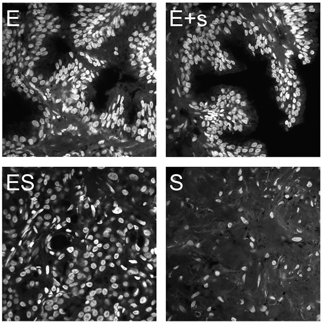
Figure 1. Prostate tissue imaging showing optical mid-sections in the DAPI-channel.

Imaging was performed to acquire four categories of tissue frames corresponding to a sampling spectrum of epithelial and stromal compartments: epithelia only (E), epithelia with minor bordering stroma (E+s), mixed epithelia and stroma at various ratios (ES), and stroma only (S).
