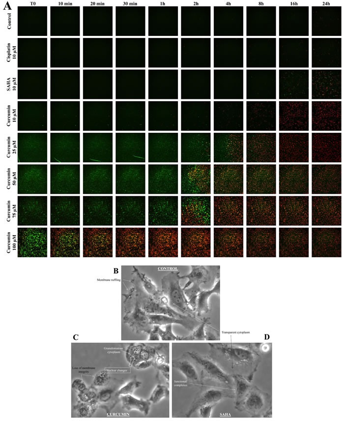
Figure 3. Timelapse fluorescence videomicroscopy of M5-T1 cells after treatment with cisplatin, SAHA or curcumin in vitro.

A., M5-T1 cells were imaged at T0, 10 min, 30 min, 1 h, 2 h, 4 h, 8 h, 16 h and 24 h after treatment with 10 μM cisplatin, 10 μM SAHA, 10 μM, 25 μM, 50 μM, 75 μM or 100 μM curcumin in comparison with control cells (incubation with normal medium containing 1% DMSO). The green fluorescent YO-PRO-1 dye and the red fluorescent propidium iodide dye were used to detect the presence of apoptotic and necrotic cells, respectively. B.-D., Morphological changes of M5-T1 cells after 2 h treatment with 100 μM curcumin in culture followed by incubation with normal medium for 3 h (C). Comparison with control cells (B) or cells treated with 10 μM SAHA (D). Changes included loss of membrane integrity and protrusion of cytoplasmic material, chromatin condensation and fragmentation, and granulomatous aspect of the cytoplasm. In contrast, junctional complexes, membrane ruffling and refringent inclusions within the cytoplasm were characteristic of living cells.
