
Figure 4. Experiment E1.
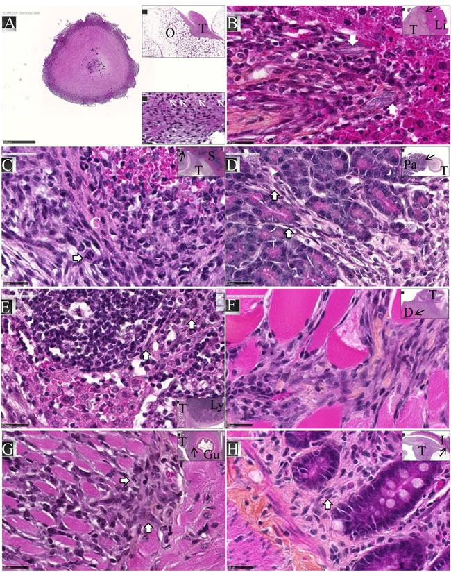
M5-T1 cells exhibit invasive capacities in vivo. HPS staining, x800, the scale bars represent 25 μm. Inserts show general views, arrows indicate the localization of magnifications, scale bars represent 500 μm. A., Example of one metastatic nodule from the peritoneal cavity, x50. The top insert shows tumor development on the omentum (O) with the formation of one of these nodules. The bottom insert exhibits a high magnification view (x800) of the area indicated by a black rectangle, with the presence of numerous monocytes/macrophages (white arrows). B., Liver metastasis (T: tumor, Li: liver). The two white arrows in the magnification show the presence of elongated tumor cells with a large nucleus moving towards the liver parenchyma. C., Spleen metastasis (S). The white arrow on the enlargement shows a mitotic figure with surrounding tumor cells invading the red pulp of the spleen after rupture of the capsule (C, in white). D., Pancreatic metastasis (Pa). The two white arrows in the magnification show the presence of elongated tumor cells moving towards the mesenchymal space separating pancreatic acini. E., Invasion of a mesenteric lymph node (Ly). Elongated tumor cells (white arrows) coming from the tumor growing in the mesentery are observed between cortical nodules close to lymphocytes. F., Invasion of the diaphragm (D), showing both clusters and elongated isolated tumor cells moving between muscle fibers. G., Invasion of the gut (Gu). The two white arrows in the magnification show the presence of tumor cells infiltrated between the longitudinal muscle cells and starting to invade the circular muscle cells of the muscularis externa. H., Metastatic tumor nodule attached to the intestine I. The white arrow indicates the presence of elongated tumor cells invading the lamina propria of the mucosa. On the left part of the figure, tumor cells are also present within the outer longitudinal layer of the muscularis mucosae.