
Figure 5. Experiment E1.
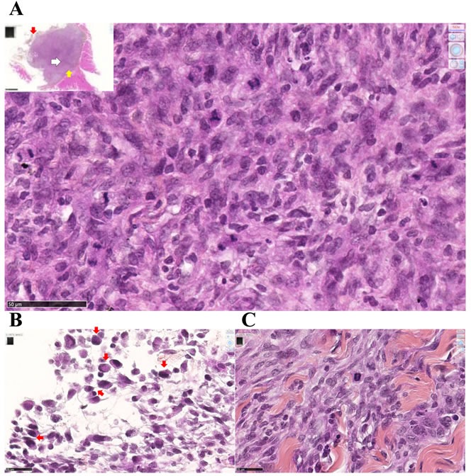
Metastatic M5-T1 tumor tissue presents a high mitotic index. Histological aspects of a representative example of metastatic mesothelioma tissue invading the parietal peritoneum, HPS staining. (A), This image corresponds to areas with the highest cell density and mitotic index observed within the tumor tissue, x600. The insert shows a general view of the tumor tissue attached to the peritoneum, the position of this area being indicated with a white arrow, x50. (B), Peritoneal cavity side, the external layer of the tumor (position indicated with the red arrow in the insert in (A)) is characterized by the presence of macrophages (red arrows), x800. (C), On the parietal side (position indicated with the yellow arrow in the insert in (A)), the interface between the tumor and the invaded peritoneum is characterized by extended areas of fibrosis, x800.