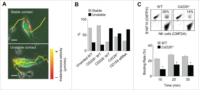
Figure 6.
Stability of contacts between NK and cancer cells. (A) Unstained NK cells (unsorted WT, CD226+ WT, CD226− WT, or Cd226−/− NK cells) and calcein AM–stained B16F10 cells (transfected with CD155 siRNA or not) were imaged every 2 min from 1 h to 5 h. Representative images of stable and unstable contacts (magnification, 200 ×; electronically zoomed; scale bar, 15 μm; n = 10 movies of 3 independent experiments per group). (B) Ratios of stable and unstable contacts between cancer cells and NK cells (n = 142, 84, 116, 111, and 146 from the left). (C) Binding rates of cancer cells and wild-type or Cd226−/− NK cells analyzed by flow cytometry (n = 3, mean ± SEM, *p < 0.01).

