
Figure 3.
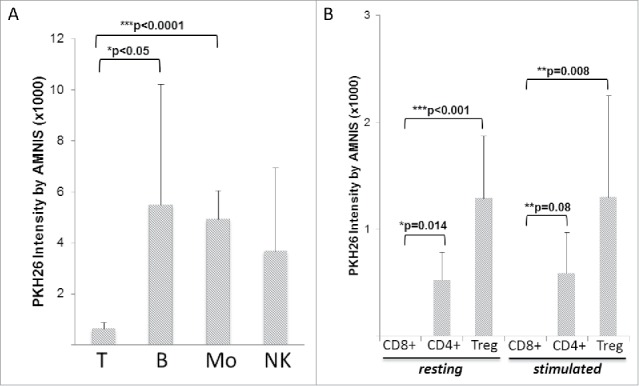
Amnis-generated data showing differences in TEX uptake by various human MNC subsets. (A) Various MNC subsets isolated from different ND were co-incubated with TEX for 24–48 h. T cells T show significantly lower TEX uptake compared with other MNC subsets. (B) Comparisons of TEX uptake by resting or activated CD8+ T cells, CD4+ T cells, or CD4+CD39+ Treg show a lack of TEX internalization by CD8+ T cells relative to low but significantly increased TEX uptake by Treg. T cell subsets were isolated from the peripheral blood of four different ND. In (A) and (B), the data are presented as mean levels of PKH26 intensity in recipient cells ± SD. The p values denote significant differences.