
Figure 4.
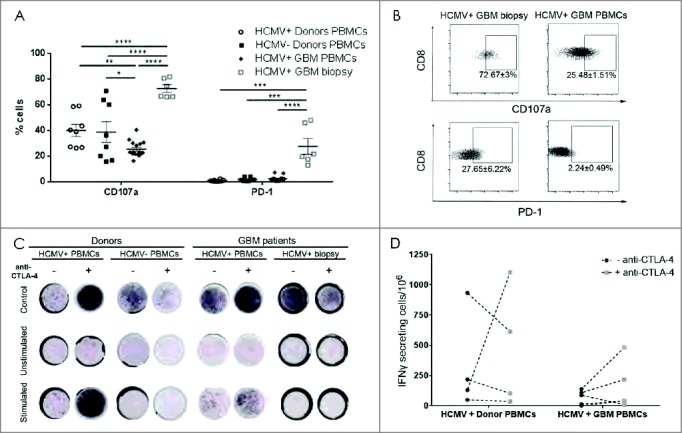
High CD107a degranulation and PD-1 expression in CD8 (T) cell from HCMV+ patient biopsies. (A) % mean ± SEM of CD107a and PD-1 expression in HCMV+ and HCMV− patients' (n = 8), tumor (n = 6) and healthy donors' (n = 8) CD3+CD8+ T cells. (B) Representative dotplots and % mean ± SEM of CD3+CD8+ T cell expressing CD107a cells and PD-1 cells. (C) Representative IFNγ ELISpots from seropositive vs. seronegative patients' blood, tumor and healthy donors with and without stimulation with pp65 or IE-1 HCMV peptides and with (+) or without (-) anti-CTLA-4 blockade. Secretion was considered positive when number of cells secreting was >0, normalized to HCMV− donors. (D) Difference in frequency of IFNγ secreting cells after stimulation with pp65 HCMV peptides in HCMV+ patients (n = 5) vs. HCMV+ donors (n = 4) with and without blocking with CTLA-4. Two way ANOVA, Bonferroni's multiple comparison, *P < 0.05, **P < 0.01, ***P < 0.001 and ****P < 0.0001.