
Figure 2.
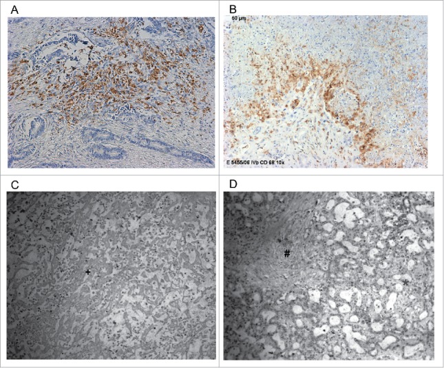
(A) Intrahepatic cholangiocarcinoma stained with mAb CD68 PG-M1 with a high abundance of TAMs in tumor central area (TCA). Original magnification: x 200. (B) Intrahepatic cholangiocarcinoma stained with mAb CD68 PG-M1 with a high abundance of TAMs in tumor-infiltrating front (TIF). Original magnification: x 200. (C, D) Histological tumor specimen with (C) necrosis and (D) fibrosis. *Vital tumor cell formations, +Tumor necrosis, #Tumor fibrosis.