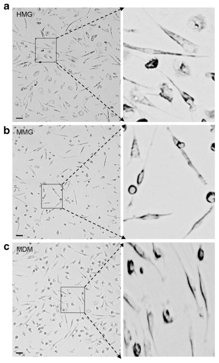
Fig. 1.
Phase contrast images of monocyte-derived microglia (MMG) and human fetal brain-derived microglia (HMG) cells. a MMG cells were generated in vitro by culturing CD14+ cells in the presence of macrophage colony-stimulating factor (MCSF), granulocyte macrophage colony-stimulating factor (GMCSF), beta-nerve growth factor (NGF-β), and CCL2 for 10–12 days. b HMG cells were isolated from 120- to 145–day-old fetal brain and cultured in high-glucose DMEM supplemented with 10 % AB-human and M-CSF for 10–12 days. c MDM were generated in vitro by culturing CD14+ cells in the presence of macrophage colony-stimulating factor (MCSF). Enlarged view of each cell type is presented on the right. Representative images of MDM, MMG, and HMG cells derived using monocytes from three independent healthy human donor bloods and fetal brain tissues, respectively. Scale bar indicates 10 μM

