
Fig. 6.
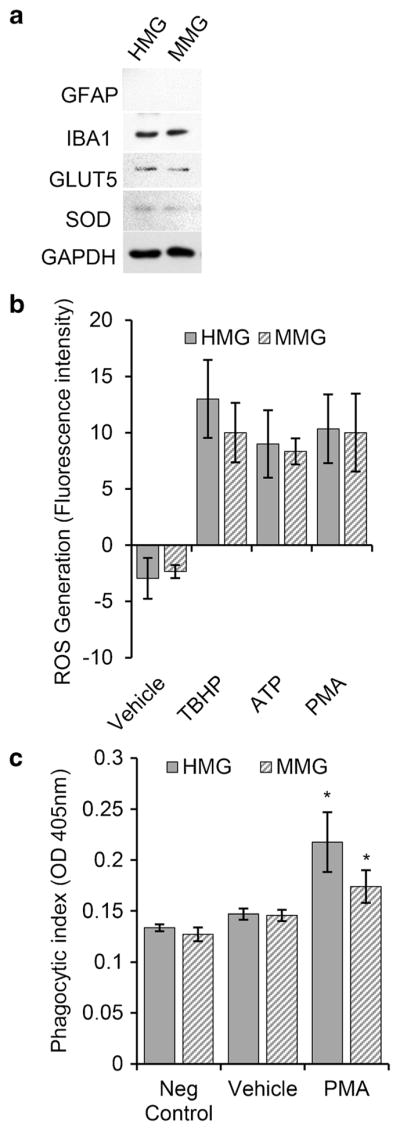
MMG and HMG cells are functional in culture. a Western blot analysis of the protein levels of different glial cell markers. MMG and HMG cells were cultured in vitro for 10 days. At day 10, cells were lysed and analyzed for endogenous glial fibrillary acidic protein (GFAP), IBA1, glucose transporter type 5 (GLUT5), superoxide dismutase (SOD), and GAPDH. b MMG and HMG cells produce ROS upon stimulation. ROS generation was determined by detecting the conversion of H2DCFDA into DCF with a fluorescence plate reader. Unstimulated MMG and HMG cells were compared with the protein kinase C activator, phorbol-myristate-acetate (PMA, 50 nM) and ATP (100 μM) stimulated cells. Tert-butyl hydrogen peroxide (TBHP, 50 μM) was used as a positive control. c Quantification of phagocytic response in MMG and HMG cells. The phagocytic ability of MMG and HMG cells in response to PMA activation was measured using prelabled zymosan (Saccharomyces cerevisiae) particles. The engulfed particles were detected using a colorimetric 96-well assay