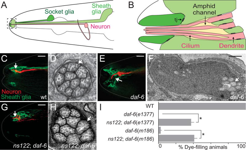
Fig. 1. ns122 is a daf-6 suppressor.

(A) The C. elegans amphid consists of 12 amphid neurons (one is shown in red) and the sheath (light green) and socket glia (dark green). The amphid channel is outlined and shown in greater detail in (B). Adapted from (Perkins et al., 1986). Tj, tight junctions. (C, E, G) The amphid channel (white arrow) in wild-type (C), daf-6(e1377) (E), and ns122; daf-6(e1377) (G) adult animals. The ASER neuron is visualized with mCherry (red, driven by the gcy-5 promoter) and the sheath glia with GFP (green, T02B11.3 promoter). Left is anterior. Scale bar, 5 μm.
(D, F, H) Transmission electron micrographs of cross-sections through the amphid channel in wild-type (D), daf-6(e1377) (F), and ns122; daf-6(e1377) (H) adult animals. White arrows point to cilia. Scale bar, 0.5 μm. Note the difference in scale between (C) and (E). (I) ns122 suppresses daf-6 dye-filling defects. n ≥ 100. Error bars, standard error of the mean (SEM), from ≥ 3 experiments. *p < 0.0001.
