Fig. 4.
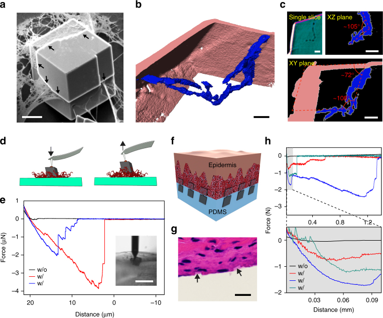
Calcite matrix as inorganic localized adhesions for biological tissues. a, b A calcite crystal incorporating collagen fibrils. a SEM image. Scale bar, 10 μm. b, c 3D reconstructed transmission X-ray microscope image b, and a single slice, and the projection images over XZ and XY planes c. Scale bars, 2 μm. d, e Schematic representation d and representative force-distance recordings e of the AFM studies of the interaction between single calcite crystals and collagen coating. (inset) Optical micrograph taken during one measurement. The AFM tip with a glue droplet first moved downward until it contacted the top facet of a calcite crystal. The force was only recorded during the retraction of AFM tip/calcite. ‘w/o’ denotes without collagen incorporation, and vice versa. Scale bar, 20 μm. f–h Flexible tissue adhesives with inorganic localized adhesions, demonstrated with a schematic f, H&E staining image g and force-distance recordings h. Scale bar, 10 μm

