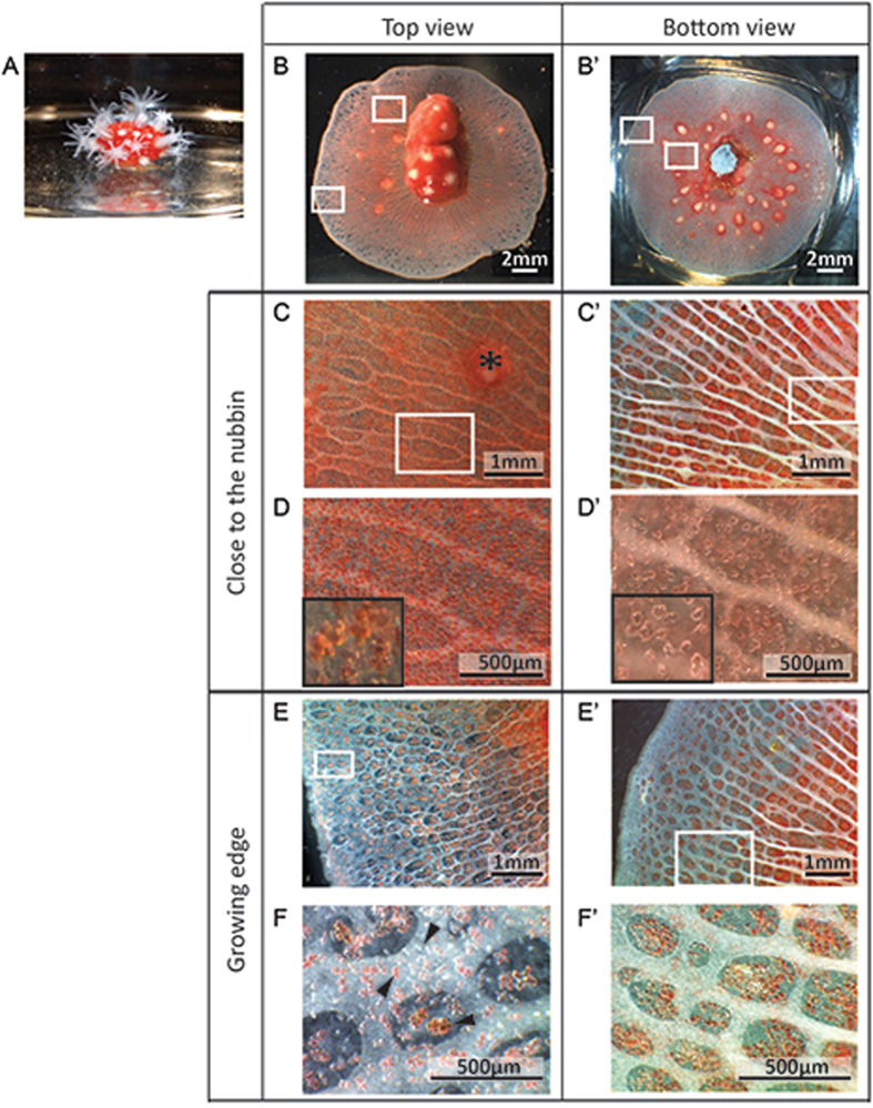
Figure 1.
Tissue organization of a representative C. rubrum microcolony. (A) A branch apex cut and glued on a glass coverslip constituting a nubbin with open polyps. (B) and (B’) General views of microcolonies with the nubbin in the center of the preparation, tissues growing around and development of polyps. Boxes indicate two regions of interest: close to the nubbin and at the growing edge. (B–F) Views from above the microcolony. (B’–F’). Views from underneath. (C) and (C’) Are enlargements of tissue close to the nubbin, the coenenchyme is red colored and contains numerous parallel white gastrodermal canals. The asterisk indicates a retracted polyp. (D) Enlargement of (C) showing the high density of red sclerites. (D’) Enlargement of (C’) showing the mineralized sheets deposited on the coverslip under tissues. (E) and (E’) Are enlargements of tissue at the growing edge showing that canals are not fully developed at the extremities and sclerites are less dense. (F) Enlargement of (E) with black arrowheads showing white, pink/orange and red sclerites. (F’) Enlargement of (E’) showing the absence of crystals deposited on the coverslip at the growing edge.

