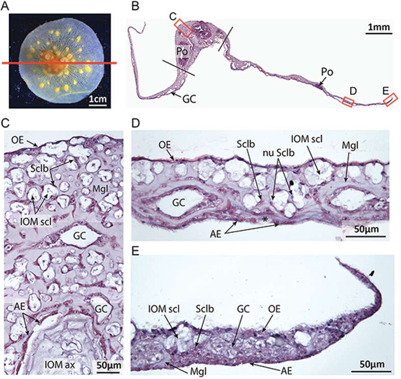
Figure 2.
Sagittal section of a decalcified microcolony stained with hematoxylin and eosin. (A) Photo of a decalcified microcolony used for sagittal section as indicated by the red line. (B) Reconstruction of the general view of the stained sagittal section with hematoxylin and eosin. The nubbin is localized between the two black lines. The adjacent parts correspond to the tissue growing on the coverslip (Scale: 1 mm). (C) Histology at the nubbin’s apex (Scale: 50 μm). (D) Histology of the tissue growing on the coverslip. The asterisk indicates secreted IOM by the aboral ectoderm (Scale: 50 μm). (E) Histology of the tissue growing at the growing edge on the coverslip (Scale: 50 μm). Po: Polyp; GC: Gastrodermal canals; OE: Oral ectoderm; AE: Aboral ectoderm; Mgl: Mesoglea; Sclb: Scleroblasts; nu Sclb: Scleroblasts nuclei; IOM scl: Insoluble organic matrix of sclerites; IOM ax: Insoluble organic matrix of the axial skeleton.

