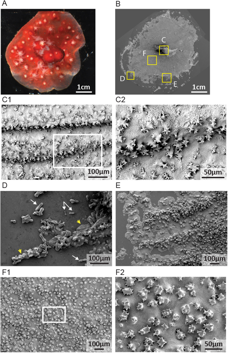
Figure 3.
Microstructure of the newly formed axial skeleton in a C. rubrum microcolony processed for SEM. White boxes indicate enlargements. (A) Microcolony before removal of soft tissues. (B) General view of crystals deposited on the coverslip. Yellow boxes indicate four regions of interest. (C1,C2) Organization of the nubbin’s axial skeleton. (D) First stage of skeleton formation with dumbbell-shaped crystals deposited (white arrows), then embedded in a mineral sheet (yellow arrows). (E) Later stage of skeleton formation with mineral sheets covered with microprotuberances. (F1,F2) More advanced stage of skeleton formation with a thick skeleton covered with a high density of microprotuberances.

