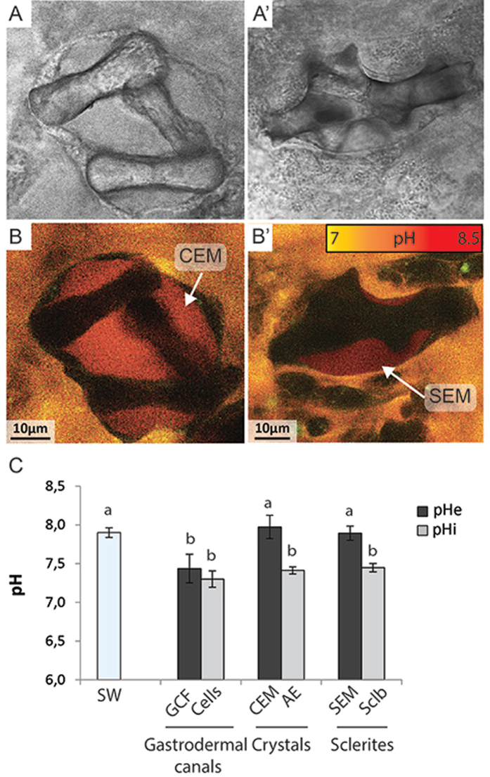
Figure 6.
Confocal images of SNARF-1 labeling of the calcifying medium of biominerals and pH measurements in different intra- and extracellular compartments. Transmitted light images of dumbbell-shaped crystals deposited on the coverslip (A) and a growing sclerite in the mesoglea (A’). (B-B’) Fluorescence images of SNARF-1 labeling showing the presence of an extracellular calcifying medium around biominerals. CEM: Cristal Extracellular Medium; SEM: Sclerite Extracellular Medium. (C) In vivo pH measurements obtained in cells and calcifying extracellular medium of C. rubrum microcolonies. Data are compared to the seawater pH (SW) surrounding microcolonies. Data are mean per microcolony ± standard deviation. SW: Seawater (n = 7); GCF: Fluid of gastrodermal canals (n = 4); CEM: Crystals extracellular medium (n = 5); AE: Aboral epithelium (n = 4); SEM: Sclerites extracellular medium (n = 4); Sclb: Scleroblasts (n = 7). Letters indicate mean pH values significantly different (one-way ANOVA with Leven’s post-hoc).

